How to prevent goldfish from parasites
Goldfish parasitic disease is one of the most serious diseases that harm goldfish. Now there are three common parasites of goldfish: fish gills, third-generation worms and anchor head lice. Teacher Zhang suggested:
1. Adult parasites can already be seen in fish tanks. Try to catch them with tweezers and apply some red mercury to the wounds of goldfish.
2. Parasites are already present in fish tanks, and goldfish are to be transferred to a healthy water environment in a timely manner. Let the parasite find no parasitic host, and the parasite will die within a few days. Thoroughly clean the tank and remove the parasites.
3, you can also try to put 90% of crystal trichlorfon 0.25-0.3 grams per cubic meter of water in the fish tank standard drug delivery, or in the 10kg water 90% crystal trichlorfon 0.5-1.0 grams, bath sick fish 10- 15 minutes, continuous medication 3-4 times.
4, can also use 0.5 grams of potassium permanganate dissolved in 50kg of water, bath sick fish for 30-60 minutes, back into the pool of water (cylinder).
Of course, the standard of water quality should be monitored at all times during the breeding process, and the water should be changed regularly to keep the water environment clean. At the same time, it is necessary to regularly sterilize goldfish to ensure the safety of the water environment, provide a good living environment for goldfish, and prevent the infection of parasites.
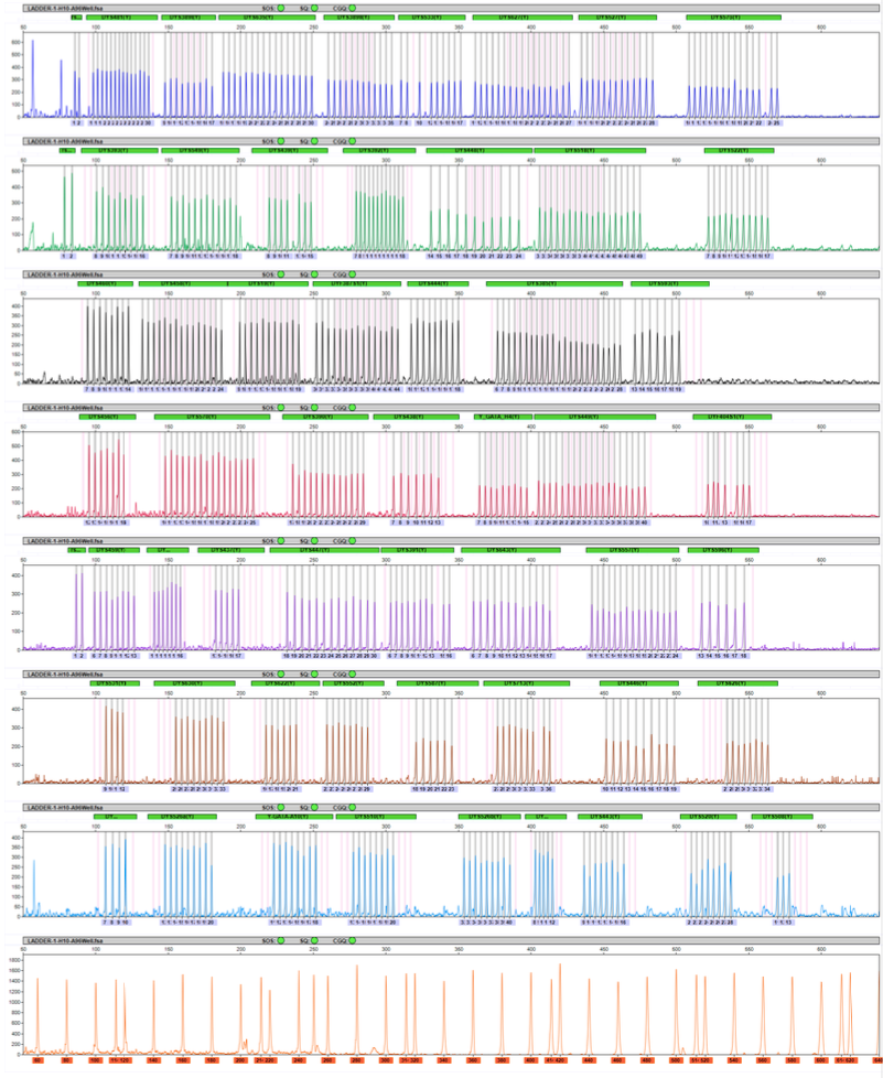
To extract a mixture of DNA fragments, put through a PCR instrument to do a simple purification. Remove the free fluorescent ddNTP single nucleotide, leaving a DNA fragment of a certain length, which can be sequenced on the machine. A polyacrylamide solution is first injected into a hollow capillary during the sequencing process. Then, the polyacrylamide solution was ionized by UV light irradiation to generate a polymerization reaction. The polyacrylamide gel produces a separation effect under the electric field to start the electrophoresis of nucleotide. The electrophoretic movement of short DNA fragments is fast, and the electrophoretic movement of long DNA fragments is slow. The mixture of DNA fragments moves from a negative charge to a positive charge under the action of an electric field in a capillary containing a polyacrylamide gel. The positive end of the capillary is irradiated with a solid-state laser, and a spectroscopic optical sensor records the different fluorescence intensities. Each DNA fragment, when passing through the laser scanning point, has a fluorescent group on it, which will emit a specific fluorescent color.
Because in the previous polymerization reaction process, the starting point of the polymerization reaction starts from a specific primer position. Therefore, the DNA fragment that reaches the laser scanning point of electrophoresis first is the shorter fragment, so its polymerization termination position will be closer to the polymerization start position. Therefore, the fluorescent color reflects which of the bases at its 3' end is A, T, C, and G.
Conversely, the slower the electrophoresis of DNA fragments reaches the laser scanning point, the longer the DNA fragments. As a result, its termination site is farther away from the starting position of the primer. Finally, a map of four colors is obtained.

Fragment Analysis Instrument,Medical Diagnosis Clinical Analyzer,Forensic Testing Dna Sequencer,Capillary Fragment Analysis Genetic Analyzer
Nanjing Superyears Gene Technology Co., Ltd. , https://www.superyearsglobal.com