Estimation of Molecular Weight of Protein by ScanLater Immunoblot Protein Ladder
2023-02-14 10:06:10
Application of ScanLater immunoblot protein ladder
Estimating the molecular weight of a protein
advantage:
· A simple set of standards for gel visualization, membrane positioning and time-resolved fluorescence detection 
· Proteins have been labeled with biotin 
· Can detect the molecular weight of the analyte 
· No need to mix and heat
Introduction
ScanLater immunoblot protein ladder is an essential component of the ScanLater immunoblot protein detection system. The protein ladder is initially applied to the quantification of protein molecules, visualization of gel electrophoresis, and the process of transmembrane has been evaluated.
The ScanLater immunoblot protein ladder consists of 7 biotinylated recombinant proteins and 3 pre-stained protein markers. The experiment detects the biotin-labeled protein by adding tritiated streptavidin to the secondary antibody stage. The labeled membrane can be read by a ScanLater immunoblot protein assay cartridge mounted on a SpectraMax I3 or SpectraMax Paradigm multi-plate reader.
ScanLater immunoblot protein ladder: The ScanLater immunoblot protein ladder contains seven biotinylated recombinant proteins with molecular weights of 10, 20, 40, 50, 80, 100 and 140 KD, respectively. They were detected by incubating the membrane with tritiated streptavidin and displayed in bright color bands on a ScanLater immunoblot protein detection system (Figure 1, left). In addition, there are three visible pre-dyed markers, 18, 31 and 70KD (Figure 1, right). These blue bands are clearly visible on the electrophoresis strip after the film is transferred. They also exhibit dark bands on the ScanLater immunoblot protein detection system. We compared a set of reference standard curves by the experiments shown in Figure 2 to determine the linear relationship between the molecular weight of biotinylated ladder proteins and their migration distance on the strip.
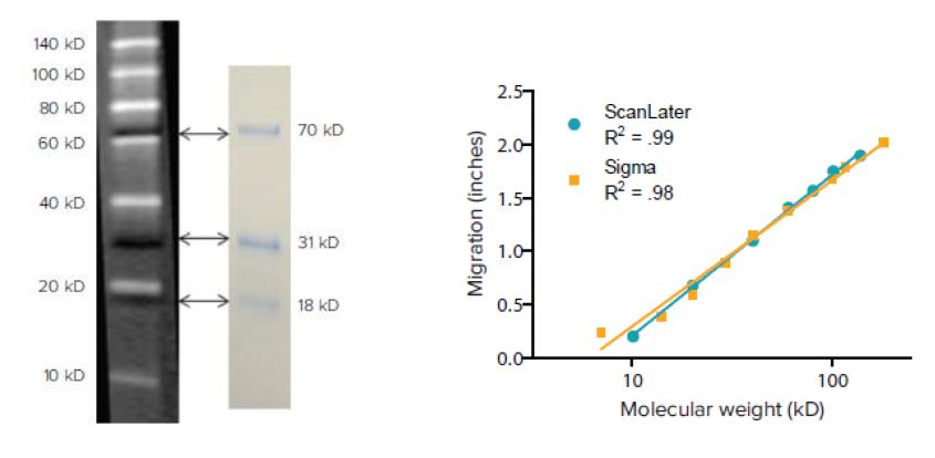
Figure 1: ScanLater immunoblot protein ladder. The white band indicates the biotinylated protein used to detect the molecular weight of the protein on the membrane. After incubation with tritiated streptavidin, the ScanLater immunoblot protein detection system can be used for reading. The dark bands are pre-stained proteins to monitor protein electrophoresis, hybridization mapping, and transfer efficiency. In these processes, these blue bands are visible to the naked eye.
Figure 2 : Migration vs molecular weight. The migration distances of the seven proteins on the ScanLater immunoblot protein ladder were plotted and fitted as a function of the respective molecular weights. The linear relationship is similar to the results of the reference protein (Cat. No. #B2787) purchased from Sigma.
ScanLater immunoblot protein detection method
The procedure for ScanLater Western Blot Protein Detection was performed with reference to standard run-through and transfer methods prior to incubation with secondary antibodies. Then, along with the target protein sample, 4 ul of ScanLater immunoblot protein ladder was added to one column of the strip. No additional heating or other preparation steps are required. After incubation with the primary antibody, blocking and washing were performed, and then both the labeled streptavidin and the europium-labeled secondary antibody were incubated at 1:5000. The é“•-labeled streptavidin binds to the protein ladder, and then the é“•-labeled secondary antibody specifically binds to the primary antibody of the target protein. Labeled membranes can be read by the ScanLater Immunoblot Protein Detection Cartridge mounted on a SpectraMax I3 or SpectraMax Paradigm multi-label plate reader that utilizes time-resolved fluorescence (TRF) to detect strontium elements. Time-resolved fluorescence detection can significantly reduce the background noise of autofluorescence, or other emitted light with a short half-life. It also effectively avoids the over-exposure that often occurs in chemiluminescence and conventional fluorescence detection; therefore, this system can provide sharp bands with high signal-to-noise ratio. Since this method does not involve an enzymatic reaction, the inherent variability in protein quantification is eliminated. In addition, since the ruthenium element has an anti-photobleaching effect, the signal on the film can be maintained for several weeks to one month and can be read repeatedly. The ScanLater Western Blot Protein Detection System, including the ScanLater Immunoblot Protein Ladder, provides a simple, sensitive and stable assay platform that provides excellent protein analysis capabilities for multi-function microplate readers.
Visual inspection of unknown proteins
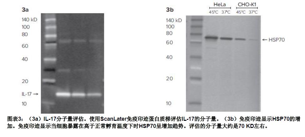
The following two experiments to detect interleukin-17 and heat shock protein, respectively, provide a good basis for the ScanLater immunoblot protein ladder to evaluate protein molecular weight.
IL-17
Interleukin-17 (IL-17) was loaded for 30 minutes in a 4-20% gradient gel in 1X buffer. The ScanLater immunoblot protein ladder was also placed on the same gel. The protein will be transferred to a fluorescent membrane and incubated with anti-rat IL-17 overnight, then 1:5000 ScanLater(R)-labeled anti-rat IgG and 1:5000 ScanLater(R)-labeled anti-streptavidin antibody. As shown in Figure 3a, when the membrane is washed and air-dried, it can be read by the ScanLater Western Blot Protein Detection Cartridge mounted on the SpectraMax I3 Multi-Function Microplate Reader. The molecular weight of IL-17 was determined to be 14 KD by ScanLater immunoblot protein ladder, which is similar to the actually published molecular weight of 15 KD.
Heat shock protein
Heat shock protein (HSP70) is induced when cells are exposed to temperatures above normal incubation temperatures. In this study, we exposed CHO-K1 cells and Hela cells to 45-90 minutes at 45 degrees Celsius, and then collected cell extracts after 6 hours. Finally, 5 ul of the extract was applied to the 4-20% gradient gel for electrophoresis, and the 4 ul ScanLater immunoblot protein ladder was also placed on the same gel. After transfer, the membrane was blocked, washed, and incubated with anti-mouse HSP70 overnight, and then sputum labeled anti-mouse IgG was incubated for 1 hour. In this experiment, the protein ladder was incubated separately from the anti-neutral marker against streptavidin. As shown in the reverse view of Figure 3b, the pre-stained standard showed an off-white band because the stain quenched the time-resolved fluorescent signal. Both cell lines showed that HSP70 increased with increasing temperature. According to the ScanLater immunoblot protein ladder, the molecular weight of the protein HSP70 was approximately 70 KD.
to sum up
The ScanLater immunoblot protein ladder allows for the evaluation of molecular size with bright bands (Figure 1), while providing visible indicators for gel electrophoresis, transfer and transfer. This system allows users to use electrophoresis and immunoblotting procedures optimized for their application. It is recommended to use a protease inhibitor that targets the anti-streptavidin and sputum-labeled secondary antibodies during incubation, so that when reading with the SpectraMax I3 multi-function microplate reader, the time resolution of the protein ladder and the target band can be effectively reduced. Fluorescence detection background. Images are captured using SoftMax Pro software, and the software can be integrated and exported to a custom Excel for quantitative analysis. With the introduction of the ScanLater immunoblot protein ladder, the ScanLater immunoblot protein detection system provides superior sensitivity and protein analysis capabilities for multi-function microplate readers.
Dehydrated Red Beet,Dehydrated Red Beets For Snacks,Dehydrated Red Beet Powder,Dehydrated Red Beet For Baking
Xinghua Lvwei Foods Co.,Ltd , https://www.lvweifoods.com